


FAQ’S
Breast Ultrasound FAQ’s
-
Dense tissue doesn’t obscure the image on ultrasound the same way it does on mammography, making it excellent for those who have been told they are “dense”.
We see through the tissue very clearly, in fact, we can see down to your lungs, but we won’t be scanning those! All levels of density welcome.
-
Breast ultrasound can evaluate many types of breast abnormalities. Common findings seen on breast ultrasound are cysts (simple and complex), fibroadenomas, solid masses, dilated ducts, fibrocystic changes, breast implants, abscesses, enlarged lymph nodes, and more.
-
Ultrasound is ideal for those with implants! It not only provides a clear look at the surrounding tissue (even behind the implant), but it can also identify any implant concerns such as rupture, leaks, capsular contracture, and other changes.
Any abnormal changes to the implants will be added to your radiology report.
-
We can perform ultrasound seamlessly on those who have had mastectomies, reduction mammoplasty, biopsies, and other breast surgeries-they do not affect our imaging!
-
Yes. Ultrasound uses sound waves, not radiation, so it’s completely safe for both mom and baby. Breastfeeding does not interfere with the scan!
Please Note: While breastfeeding, milk can make the ducts look temporarily enlarged on ultrasound. We will make note if this is the case.
-
While we believe ultrasound is a superior imaging modality for its safety and accuracy, especially for women with dense tissue, mammography can be best used as a compliment to sonography.
-
Yes, we scan the entirety of both breasts and the underarms (axillae) to check for any changes in the lymph nodes.
-
No, we do not currently accept insurance, which is an intentional choice, to make your experience simpler and stress-free. By offering out-of-pocket services, we eliminate the delays, referral requirements, and scheduling hurdles often associated with insurance-based care. This allows you to book your screenings quickly and on your terms!
Other Exam-Specific FAQ’s
Pelvic
-
Ovarian cysts, uterine fibroids, misplaced IUD’s, adnexal masses, free fluid, and much more. Pelvic ultrasounds are one of the best ways to visualize the uterus and ovaries clearly and in real time!
-
Yes. An ultrasound is one of the most common and effective ways to detect uterine fibroids. It can help identify the number, size, and location of fibroids. In many cases, fibroids are found during imaging even in women who do not have symptoms.
-
A pelvic ultrasound can provide detailed information about the appearance and structure of your ovaries. It can help identify common findings such as ovarian cysts, follicles, masses, PCOS, enlarged ovaries, fluid around the ovaries and more!
-
Yes, ultrasound is excellent at viewing IUD’s very clearly. If you suspect your IUD has moved down to the cervix or up toward the bladder, we can visualize where exactly it is.
-
Patients must drink 3-6 cups of water 1 hour before the appointment and cannot empty the bladder until after the exam.
A full bladder helps move the intestines out of the way, making it easier to see the uterus and ovaries.
We apply light pressure to the pelvic area, just below the belly button.
These instructions will be emailed to you after scheduling an appointment.
-
Yes, we offer transabdominal (external, on top of the pelvis) to view the uterus and ovaries.
This is safe for patients who are not sexually active and it is non-invasive.
-
Yes, the transducer glides gently on the pelvis region and is safe for women who may be on their menstrual cycle.
This exam requires a full bladder and can be a little less comfortable while menstruating.
-
Urgent care is highly recommended if you are having excruciating pain or symptoms.
Our role is to offer preventative imaging services. We do not replace a doctor’s office, clinic, or hospital.
Thyroid
-
Yes, thyroid blood tests tell us how well the thyroid is functioning, but they don’t show us what the thyroid looks like. It’s possible to have normal thyroid hormone levels while still having a thyroid nodule, cyst, enlargement, or other structural abnormality that wouldn’t necessarily affect blood work.
-
Yes, a thyroid nodule can sometimes cause symptoms if it becomes large enough or if it is positioned in a way that presses on nearby structures in the neck, sometimes causing a “bulge” or discomfort when swallowing.
-
Common symptoms from a larger thyroid nodule or thyroid enlargment include:
Feeling a lump or bulge in the front of the neck
A sensation that something is stuck in the throat
Difficulty swallowing. or discomfort swallowing
Needing to clear the throat frequently
Hoarseness or voice changes
-
Thyroid function is evaluated through blood tests that measure hormone levels. However, a thyroid ultrasound can provide valuable information about the gland’s structure, including the presence of nodules, cysts, enlargement, inflammation, or other abnormalities. When combined with lab testing, ultrasound helps provide a more complete assessment of thyroid health.
-
Yes. Enlarged lymph nodes occur for many reasons, including infections, autoimmune conditions, inflammation, and other medical concerns.
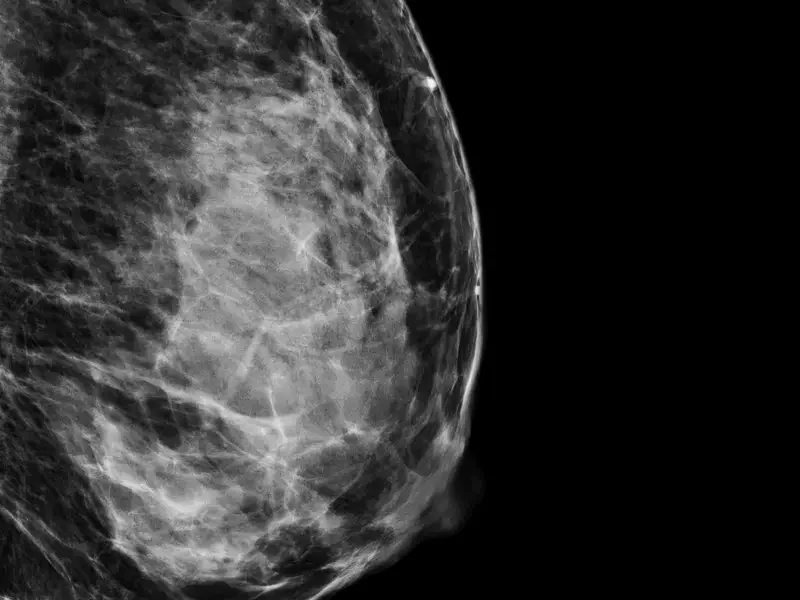
a.
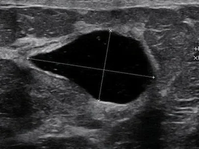
b.
Mammogram vs
Ultrasound
On a mammogram, dense breast tissue looks white (a), which can make it harder to see cysts or solid lumps. With ultrasound (b), dense tissue, cysts, and solid lumps each look different. The dense tissue doesn’t obscure the image.
The cyst is clearly seen on the ultrasound and appears as a “black” lesion, the dense tissue seen around it doesn’t block the view.
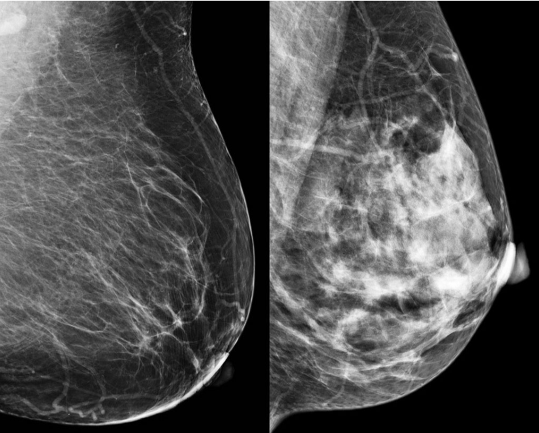
A fatty breast (left) versus a very dense breast (right) on mammography. Cysts and solid masses also appear white on mammography, making it less sensitive on women who have very dense breast tissue.
General Questions
-
No! We are an out-of-pocket facility because most insurances won’t cover ultrasound screenings. We make it easy to make an appointment, you can book online or call our office at your convenience!
-
Ultrasound uses sound waves instead of X-rays, so there’s no radiation, and it works especially well in women with dense breasts, since density doesn’t block the sound waves. It can also visualize tissue around implants and evaluate implant integrity. There is no compression, making it ideal for those with implants.
Scroll to the bottom of the page to see the difference between mammography and ultrasound
-
No, we do not currently accept insurance. This is an intentional choice to make your experience simpler and stress-free. By offering direct-pay services, we eliminate the delays and scheduling hurdles often associated with insurance-based care. This allows you to book your screenings quickly and on your terms!
-
We are looking for any abnormalities! We can see the differences between cysts, solid masses, calcifications, and more. We can even see behind breast implants.
We capture detailed images so that changes can be monitored over time, helping to ensure that nothing develops into something more concerning.
-
Patients are welcome to schedule additional imaging at any time with us to monitor areas of concern, whether for their peace of mind or at the recommendation of their healthcare provider. The radiologist will also recommend a follow-up on your report if he feels it’s necessary.
Your provider may request the DICOM images of your exam. To release records by email, please provide your physician’s contact information at the time of service, or have your provider contact our office directly at (714) 371-8047 or by email at results@radiantviewultrasound.com.
Important Note:
Your doctor still may recommend additional imaging-such as a mammogram or other tests- to complete the evaluation if abnormalities are seen. These decisions are made by your physician and are outside our control.
-
Our practice is an out of pocket service, which means we do not bill insurance and we do not provide superbills for reimbursement . We’ve chosen this model so we can keep our services simple and more affordable. We provide straightforward pricing and focus on giving you high-quality care without hidden fees.
We do, however, provide an itemized receipt which may be used for HSA and FSA reimbursements.
-
All exams are reviewed by a Board-Certified Radiologist, who provides a detailed report. Reports are typically ready within 1-3 business days. Please note that we do not have a radiologist on site.
While our ultrasound technologist is highly skilled in conducting the scan, they are not permitted to provide diagnostic information during your visit.
-
Yes — at RadiantView, we offer elastography, an advanced ultrasound technique that measures how stiff or soft tissue is. Benign findings tend to be softer, while areas of concern tend to be firmer. This extra layer of information helps physicians evaluate findings with more confidence, which can mean fewer unnecessary biopsies, follow-ups, and less worry for patients. Compared to all other ultrasound methods, including 3d, automated, and QT ultrasound, elastography cuts down on the 'gray area' guesswork by giving a clearer picture of what's actually going on. And like the rest of your ultrasound, it's completely painless and radiation-free — performed during the same visit.
We are the only screening facility in all of Orange County to carry this technology!
-
We accept major credit cards including Visa, Mastercard, American Express, Discover, and Apple Pay.
We also take FSA and HSA debit cards as long as they have a major credit card logo.
An itemized receipt will be provided which may be used for HSA and FSA reimbursements.
-
Our Highlights:
Comprehensive screenings
Get all your ultrasound needs in one place
Women’s health expertise
Specialized care from a licensed ultrasound technologist with a focus and passion in women’s health
Hospital grade technology
Top-tier machines for accurate results with advanced technology
Comfortable facility
Designed to put you at ease from start to finish! (warm gel, cozy blanket, soothing music, gentle care)
Fast results
Reports delivered within 1-3 business days by a California licensed Radiologist
Personalized care
No “assembly line” approach, we take the time needed with each patient
Patient satisfaction first
Our goal is for you to leave feeling cared for, comfortable, and at ease
-
You may log into your account to modify or reschedule your appointment at any time up to 24 hours before your scheduled visit.
Call our office directly if
-
Absolutely! Although we are not affiliated with an insurance company or hospital, your ultrasound is interpreted by a licensed board-certified radiologist, and the official report and images can be kept in your medical records just like any other imaging study.
Your provider may request the DICOM images of your exam. To release records by email, please provide your physician’s contact information at the time of service, or have your provider contact our office directly at (714) 371-8047 or by email at results@radiantviewultrasound.com. We can also fax the results upon request.
-
Insurance usually only covers ultrasounds when they’re medically necessary—meaning a doctor has ordered the test to diagnose or monitor a specific health concern.
Screenings, on the other hand, are preventative and done proactively to check on your health before symptoms appear. Since they aren’t tied to a diagnosis or treatment plan, most insurance companies don’t consider them “essential” and won’t cover the cost.
That’s why we offer our services directly to you-no referral, no long wait, and no surprise billing. Just easy, transparent access to care.
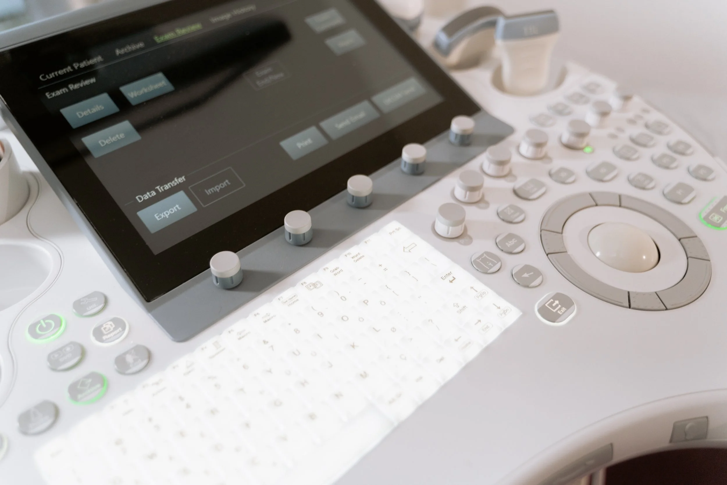
All exams include a detailed Radiologist’s report - sent directly to you within 1-3 business days. No insurance involvement means faster care, and better technology means more reassurance!